AG Groß
Extracellular Signal Transduction
Research
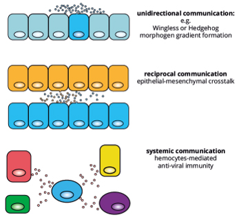
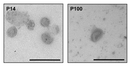
The research focuses on the biogenesis of extracellular vesicles (EVs) and their role in Wnt protein secretion, particularly in Drosophila models and cancer cells. EVs are membrane-bound vesicles secreted by all types of cells, which transfer proteins, RNA, and lipids to other cells. This has been key in identifying how EVs form and influence cellular processes in both health and diseases.
In EV biogenesis, a critical finding was the role of neutral sphingomyelinases in EV formation at MVBs but also the plasma membrane. These enzymes are central to the budding and release of EVs into the extracellular space, affecting the number and type of signaling molecules they carry. Sphingomyelinase activity thus governs EV production, which in turn impacts various signaling pathways, helping maintain cellular equilibrium and enabling efficient cell-to-cell communication - especially relevant in pathological conditions like cancer.
Wnt signaling, another major focus of the group, is intricately linked with EVs. In Drosophila, studies reveal that Wnt proteins are frequently transported on exosomes or sEV, allowing these lipid-modified proteins to travel through the extracellular matrix and reach target cells. This EV-based Wnt transport is crucial for proper tissue development and cell differentiation, illustrating the evolutionary conservation of Wnt signaling mechanisms. Drosophila is an excellent model to study protein function in the cellular stress context, as 75% of disesase-related proteins are evolutionary conserved from fly to human.
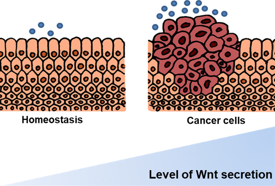
In cancer, Wnt proteins within EVs play a role in tumor progression and metastasis by enhancing intercellular communication within the tumor environment. Research demonstrates that macrophage-derived EVs carrying Wnt5a promote invasive behavior in cancer cells. By delivering oncogenic signals directly to malignant cells, EVs contribute to the cancer’s metastatic potential, highlighting them as promising therapeutic targets. Disrupting EV production or blocking specific cargo delivery may offer new approaches to interfere with cancer progression.
This work collectively advances our understanding of EV-mediated Wnt signaling in both development and disease, suggesting potential diagnostic appliations that target EV formation or manipulate their cargo to inhibit disease-related signaling pathways.
Current Projects
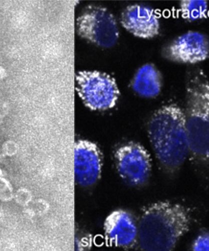
Neutral sphingomyelinases facilitate EV budding from the plasma membrane and into multivesicular bodies, a key step in their biogenesis. This process underlies many cellular signaling mechanisms, including in cancer and inflammatory responses (Menck et al., 2017). The neutral sphingomyelinase project focuses on this enzyme’s role in EV formation at the plasma membrane. This enzyme regulates the release of vesicles by modifying membrane lipid composition, facilitating EV budding and subsequent detachment from the membrane. By controlling EV production, neutral sphingomyelinase influences how cells communicate with their environment, a process that has implications for various conditions, including cancer and immune responses. This project emphasizes the role of lipid-modifying enzymes in EV biogenesis and their impact on signaling dynamics in both normal and diseased tissues.

The role of EVs in Wnt protein transport and factors essential for cellular signaling in development and disease. This study found that Wnt proteins are packed onto exosomes, facilitating their extracellular spread and effect on target cells, particularly in cancer (Gross et al., 2012). The kinesin project investigates how motor proteins, specifically certain kinesins, transport Wnt proteins within cells toward MVBs, where they are packaged into exosomes. Kinesins move along microtubules, acting as directional transporters for Wnt proteins and guiding them to MVBs. This intracellular transport is crucial because it enables Wnt proteins to be secreted in a lipid-modified form, avoiding extracellular matrix barriers that would otherwise hinder their spread. This mechanism allows Wnt to establish precise signaling gradients required for developmental processes in Drosophila, such as wing formation. This project provides insights into the importance of intracellular motor proteins in controlling extracellular signaling pathways.
Our Drosophila EV model is an innovative in vivo system designed to study extracellular vesicle (EV) formation and secretion. Using tissue-specific labeling and genetic tools like RNAi, it enables visualization of EVs in different tissues and detailed biochemical analysis. Research using this model has identified Rab GTPases, including Rab11 and Rab35, as key regulators of EV secretion from the fat body, along with new roles for Rab14 and the kinesin Klp98A. This model offers significant insights into EV biology, intercellular communication, and organ development.
Group Members

Group leader: Prof. Dr. rer. nat. habil. Julia C. Groß
E-Mail: julia.gross@hmu-potsdam.de | Online Profile: Google Scholar
2024
- Welsh, J. A., Goberdhan, D. C. I., O'Driscoll, L., Buzas, E. I., Blenkiron, C., Bussolati, B., Cai, H., Di Vizio, D., Driedonks, T. A. P., Erdbrügger, U., Falcon-Perez, J. M., Fu, Q. L., Hill, A. F., Lenassi, M., Lim, S. K., Mahoney, M. G., Mohanty, S., Möller, A., Nieuwland, R., Ochiya, T., … Witwer, K. W. (2024). Minimal information for studies of extracellular vesicles (MISEV2023): From basic to advanced approaches. Journal of extracellular vesicles, 13(2), e12404. https://doi.org/10.1002/jev2.12404
2023
- Schoger, E., Bleckwedel, F., Germena, G., Rocha, C., Tucholla, P., Sobitov, I., Möbius, W., Sitte, M., Lenz, C., Samak, M., Hinkel, R., Varga, Z. V., Giricz, Z., Salinas, G., Gross, J. C., & Zelarayán, L. C. (2023). Single-cell transcriptomics reveal extracellular vesicles secretion with a cardiomyocyte proteostasis signature during pathological remodeling. Communications biology, 6(1), 79. https://doi.org/10.1038/s42003-022-04402-9
2022
- Tietz, H. and Gross, J. C. (2022) Kinesin-mediated transport in the secretion of extracellular vesicles. Trillium Extracellular Vesicles 1 (5). https://doi.org/10.47184/tev.2023.01.06
- Linnemannstöns, K., Karuna M, P., Witte, L., Choezom, D., Honemann-Capito, M., Lagurin, A. S., Schmidt, C. V., Shrikhande, S., Steinmetz, L. K., Wiebke, M., Lenz, C., & Gross, J. C. (2022). Microscopic and biochemical monitoring of endosomal trafficking and extracellular vesicle secretion in an endogenous in vivo model. Journal of extracellular vesicles, 11(9), e12263. https://doi.org/10.1002/jev2.12263
- Choezom, D., & Gross, J. C. (2022). Neutral sphingomyelinase 2 controls exosome secretion by counteracting V-ATPase-mediated endosome acidification. Journal of cell science, 135(5), jcs259324. https://doi.org/10.1242/jcs.259324
- Choezom, D. & Gross J. C. (2022). Characterization of two novel neutral sphingomyelinase 2 inhibitors in endosomal sorting and extracellular vesicle biogenesis. Trillium Extracellular Vesicles. https://doi.org/10.47184/tev.2022.01.02
2021
- Verweij, F. J., Balaj, L., Boulanger, C. M., Carter, D. R. F., Compeer, E. B., D'Angelo, G., El Andaloussi, S., Goetz, J. G., Gross, J. C., Hyenne, V., Krämer-Albers, E. M., Lai, C. P., Loyer, X., Marki, A., Momma, S., Nolte-'t Hoen, E. N. M., Pegtel, D. M., Peinado, H., Raposo, G., Rilla, K., … van Niel, G. (2021). The power of imaging to understand extracellular vesicle biology in vivo. Nature methods, 18(9), 1013–1026. https://doi.org/10.1038/s41592-021-01206-3
- Witte, L., Linnemannstöns, K., Honemann-Capito, M., & Gross, J. C. (2021). Visualization and Quantitation of Wg trafficking in the Drosophila Wing Imaginal Epithelium. Bio-protocol, 11(11), e4040.
- Gross J. C. (2021). Extracellular WNTs: Trafficking, Exosomes, and Ligand-Receptor Interaction. Handbook of experimental pharmacology, 269, 29–43. https://doi.org/10.1007/164_2021_531
2020
- Karuna M, P., Witte, L., Linnemannstoens, K., Choezom, D., Danieli-Mackay, A., Honemann-Capito, M., & Gross, J. C. (2020). Phosphorylation of Ykt6 SNARE Domain Regulates Its Membrane Recruitment and Activity. Biomolecules, 10(11), 1560. https://doi.org/10.3390/biom10111560

Dr. rer. nat. Kirsten Wunderlich (Postdoc)
E-Mail: kirsten.wunderlich@hmu-potsdam.de
2024
- Pfaller, A. M., Kaplan, L., Carido, M., Grassmann, F., Díaz-Lezama, N., Ghaseminejad, F., Wunderlich, K. A., Glänzer, S., Bludau, O., Pannicke, T., Weber, B. H. F., Koch, S. F., Bonev, B., Hauck, S. M., & Grosche, A. (2024). The glucocorticoid receptor as a master regulator of the Müller cell response to diabetic conditions in mice. Journal of neuroinflammation, 21(1), 33. https://doi.org/10.1186/s12974-024-03021-x
2023
- Wunderlich, K. A. (2023). Shining light on extracellular vesicles – EVs in the retina. Trillium Extracellular Vesicles. https://doi.org/10.47184/tev.2023.01.05
2022
- Kaplan, L., Drexler, C., Pfaller, A. M., Brenna, S., Wunderlich, K. A., Dimitracopoulos, A., Merl-Pham, J., Perez, M. T., Schlötzer-Schrehardt, U., Enzmann, V., Samardzija, M., Puig, B., Fuchs, P., Franze, K., Hauck, S. M., & Grosche, A. (2023). Retinal regions shape human and murine Müller cell proteome profile and functionality. Glia, 71(2), 391–414. https://doi.org/10.1002/glia.24283
- Nagel-Wolfrum, K., Fadl, B. R., Becker, M. M., Wunderlich, K. A., Schäfer, J., Sturm, D., Fritze, J., Gür, B., Kaplan, L., Andreani, T., Goldmann, T., Brooks, M., Starostik, M. R., Lokhande, A., Apel, M., Fath, K. R., Stingl, K., Kohl, S., DeAngelis, M. M., Schlötzer-Schrehardt, U., … Wolfrum, U. (2023). Expression and subcellular localization of USH1C/harmonin in human retina provides insights into pathomechanisms and therapy. Human molecular genetics, 32(3), 431–449. https://doi.org/10.1093/hmg/ddac211
- Demais, V., Pohl, A., Wunderlich, K. A., Pfaller, A. M., Kaplan, L., Barthélémy, A., Dittrich, R., Puig, B., Giebel, B., Hauck, S. M., Pfrieger, F. W., & Grosche, A. (2022). Release of VAMP5-positive extracellular vesicles by retinal Müller glia in vivo. Journal of extracellular vesicles, 11(9), e12254. https://doi.org/10.1002/jev2.12254
- Grotz, S., Schäfer, J., Wunderlich, K. A., Ellederova, Z., Auch, H., Bähr, A., Runa-Vochozkova, P., Fadl, J., Arnold, V., Ardan, T., Veith, M., Santamaria, G., Dhom, G., Hitzl, W., Kessler, B., Eckardt, C., Klein, J., Brymova, A., Linnert, J., Kurome, M., … Klymiuk, N. (2022). Early disruption of photoreceptor cell architecture and loss of vision in a humanized pig model of usher syndromes. EMBO molecular medicine, 14(4), e14817. https://doi.org/10.15252/emmm.202114817

M.Sc. Henrike Tietz (Phd candidate (rer. med.))
E-Mail: henrike.tietz@hmu-potsdam.de
2022
- Tietz, H. and Gross, J. C. (2022) Kinesin-mediated transport in the secretion of extracellular vesicles. Trillium Extracellular Vesicles 1 (5). https://doi.org/10.47184/tev.2023.01.06
M.Sc. Elsa Kratz (Phd candidate (rer. med.))
E-Mail: elsa.kratz@hmu-potsdam.de

B.Sc. Beatriz Atocha Czaplinska (M.Sc. candidate)
E-Mail: beatriz.atocha@hmu-potsdam.de

B.Sc. Edward Kaba (M.Sc. candidate)
E-Mail: edward.kaba@uni-potsdam.de
Cand. med. Anna Bofferding (Doctoral candidate (Dr. med.))
Cand. med. Janis Lorbeer (Doctoral candidate (Dr. med.))
Cand. med. Charlott Denkmeier (Doctoral candidate (Dr. med.))
Cand. med. Jonathan Selke (Doctoral candidate (Dr. med.))
Cand. med. Sophie Pätzold (Doctoral candidate (Dr. med.))
Open Positions in the Groß Group
AG Groß is currently looking for highly motivated candidates for the following vacant positions:
Medical PhD Students/M.Sc. Students (m/f/d)
at the Medical Faculty of the HMU
Application
Send us a short e-mail about your research interest to julia.gross@hmu-potsdam.de.
HMU, Professor Dr. Julia Groß, Schiffsbauergasse Nr. 14, 14467 Potsdam